- Summary
- Symptoms
- Read More
- Video
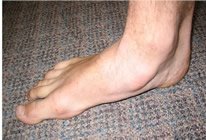
The term flatfeet (flatfoot) is used to describe a foot with a decreased or absent arch. Most cases of flatfeet are benign and never have a significant impact on the person or the activities in which they may participate during their lifetime. Some flatfeet have very specific problems that may become very painful and limit the ability to stand for any prolonged period of time. Flatfeet are found equally in men and women. Flatfeet can be congenital or acquired.
Symptoms
Congenital flatfeet (from birth):
-
May be asymptomatic or may present with pain that increases with the duration of time spent on the feet
-
Symptoms known to increase in the second and third decades of life
-
Feet may be flexible or very rigid
-
Difficulty keeping up with others for any distance or time on the feet
Acquired flatfeet
-
Begin as flexible but become rigid over time
-
Loss of arch is abrupt and often associated with an injury of the foot
-
Pain found in the medial arch, medial ankle and lateral ankle
Description
The term flatfeet is a subjective term, used to describe a foot with a decreased or absent arch. Flatfeet can be congenital or acquired. The vast majority of flatfeet are congenital. Just as we inherit facial features, eye color and hair color we also inherit a set of bones and joints that function much like those of our parents and grandparents. Most cases of flatfeet are benign, inherited and never have a significant impact on the person.
Pediatric Flatfeet
 Pediatric flatfeet are a common complaint seen in the podiatrist's office. Children will often give indirect clues of a problem like symptomatic flatfeet. They'll ask to be carried or they'll want their legs and feet to be rubbed. In the case of symptomatic flatfeet, children will tend to express these complaints more so after they've been active. The symptoms of pediatric flatfoot are due to the fact that the flatfoot is biomechanically inefficient. Simply put, it takes more work to walk with a flat foot. Therefore, kids with flatfeet have to exert more effort to keep up with other kids.
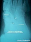
Although most cases of flatfeet are benign, there is a specific type of flatfoot that causes pain and can be of significant concern. Tarsal coalition is a foot problem that results in rigid, painful flatfoot. Tarsal refers to the bones of the rearfoot, and coalition refers to a bridge between those bones. Tarsal coalition describes a coalition or bridge of bone that forms between two bones, limiting the range of motion of the joints of the foot. The end result is a rigid, painful flatfoot. Tarsal coalition is a challenging condition to diagnose in young children. The challenge lies in the fact that the radiographic findings of tarsal coalition don't become evident until the late teens. In young children, the bridge of bone is made of fibrous material and cannot be seen in an X-ray. As the patient matures, the fibrous bridge begins to ossify, making the foot rigid.
Adult Flatfeet
Adults with inherited flatfeet can have many of the same problems as those we've discussed in children. Most adults simply complain of fatigue and an inability to get through the day comfortably. These are the same kids we've just talked about, only they've grown up.
There are specific clinical problems that cause flatfoot in the adult. Acquired flatfeet in adults is often due to damage to the posterior tibial tendon resulting in a condition known as posterior tibial tendon dysfunction (PTTD.) PTTD is the result of a partial or complete tear of the posterior tibial tendon. PTTD is a progressive condition that often results in a rigid flatfoot.
Causes and Contributing Factors
The single most common contributing factor to flatfeet is equinus. Equinus refers to a tight calf and Achilles tendon that results in limitation of the normal range of motion of the ankle. Equinus is a deforming force applied to the foot from the time a child begins to walk. If equinus is present and the arch of a young child is weak, the arch will collapse or deform under the biomechanical load generated by the calf. The collapse of the arch due to equinus provides a small amount of dorsiflexion of the foot, helping the toes move toward the shin and enabling normal walking. If equinus is significant and the arch is strong and resilient, the child will be a toe walker.
Differential Diagnosis
The differential diagnosis for flatfoot includes:
Arthritis
Charcot joint
Equinus
Fracture of the navicular
Posterior tibial tendon dysfunction
Tarsal coalition
Treatment
Examining and treating flatfeet is, to a great degree, a subjective process, but a few clinical tests are used regularly when assessing the pediatric and adult flatfoot. The clinical exam focuses on two main categories of care: is this a pediatric or adult flatfoot and is this a flexible or rigid flatfoot. 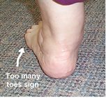 Non-weight bearing and weight bearing comparisons of the arch are always made. Toe raises are used to test tendon function. Pain in the medial arch while raising up on the toes may be suggestive of PTTD. The "too many toes" sign is a test used to evaluate abduction of the forefoot (transverse plane deformity.) Equinus is assessed by measuring range of motion of the ankle. X-rays are helpful in determining the central zone of collapse of the arch, often referred to as the midfoot fault. X-rays are also helpful in determining abduction of the forefoot as seen clinically with the "too many toes" sign.
Treatment of the pediatric and adult flatfoot focuses on management of pain. Conservative care begins with simple support of the arch. Treatment of pediatric flatfeet starts with pediatric arch supports (UCBL) and shoe modifications.(1,2) Arch supports can be over-the-counter or prescription. Shoe modification can be performed by your pedorthist or shoe repair shop and can include an arch cookie and reverse Thomas heel. A traditional oxford-style shoe is the most common shoe that can accommodate these modifications. Prescription orthotics are the mainstay of conservative care for adult flatfoot.(3,4)
The key to initial treatment of pediatric flatfeet is to try arch supports and shoe modifications to see how well they work. How do you know that they're working? You'll see a decrease in symptoms. The other consideration with kids is that they're going to grow out of things quickly. It's money well-spent to discuss your concerns with your podiatrist or pedorthist. They can recommend a treatment plan that may be significantly more cost-effective for your child in the long run.
Initial treatment of adult flatfeet follows much the same pathway as the treatment of pediatric flatfeet. Over-the-counter (OTC) arch supports are very effective in managing most adult flatfoot symptoms. Prescription orthotics are another popular method of care. Symptomatic flatfeet that do not respond to OTC or Rx inserts should be evaluated for specific problems such as PTTD.
Surgery
When a patient is evaluated for flatfoot surgery the first consideration is whether the foot is flexible or rigid.(5,6) Determining flexibility versus rigidity is subjective. Your doctor will manipulate the foot to determine its flexibility or rigidity. This determination is important in defining the surgical treatment plan. Flexibility is assessed in all three cardinal planes: frontal, transverse and sagital. Flexible flatfeet can be treated with procedures that are ambulatory, with little post-operative disability. Rigid flatfeet, on the other hand, require a higher intensity of care with a subsequently longer period of post-op care.
Flexible flatfeet
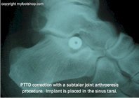 One procedure used to treat flexible flatfeet involves placing a small metal implant in the subtalar joint to wedge the foot and ankle into a more stable position. This procedure is referred to as a subtalar arthroeresis (7-14). Arthroeresis is not as invasive as other forms of surgical arch reconstruction, but may only be used in select cases of flexible flatfeet. Subtalar arthroeresis is often referred to as an internal cast, supplying support from within the subtalar joint. Subtalar arthroeresis is often performed with a procedure to lengthen the calf muscle and/or Achilles tendon. These procedures include an endoscopic gastrocnemius recession and/or Achilles tendon lengthening.
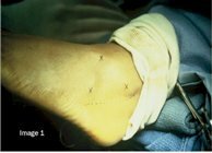
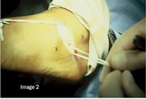
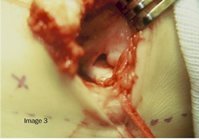
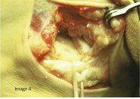
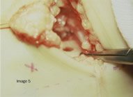
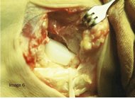
The following images show the steps used to perform an STA-Peg procedure as described by Lepow and Smith in 1989.(14) The STA-Peg procedure was one of the earliest methods of subtalar arthroeresis. Image 1 shows pre-operative planning marking the boundaries of the peroneal tendons and intermediate dorsal cutaneous nerve. In Image 2, we see the peroneal tendons retracted down and the intermediate dorsal cutaneous nerve retracted up. Image 3 shows entry into the subtalar joint. Images 4 and 5 show preparation of the subtalar joint for the implant. Image 6 shows the implant in place. Patients can bear weight on the foot the same day. STA-Peg implants come in three sizes. Image 7 shows the implants and their corresponding insertion and sizing tools.
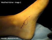
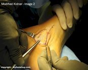
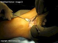
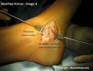
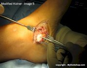
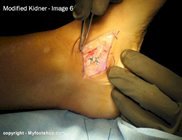
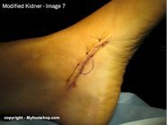
The next flatfoot procedure we'll describe is called a modified Kidner procedure. A modified Kidner procedure is commonly used in cases of flexible pediatric flatfoot. The pictures below show the steps of the procedure. A modified Kidner procedure often is performed with other procedures to correct a flexible flatfoot deformity. The procedure is also used in cases of a symptomatic os tibial externum or accessory bone of the medial arch as seen in the image to the left. Image 1 shows the planned approach with the leg to the left and toes to the upper right. Image 2 shows deep tissue dissection and identification of the posterior tibial tendon sheath. Images 3, 4, and 5 show dissection of the os tibiale externum from its investment from within the posterior tibial tendon. Image 6 shows repair of the posterior tibial tendon with non-absorbable suture. Image 7 is final skin closure.
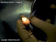
Image 8 shows the articular surface of a large os tibial externum. Os tibiale externum is found in 15 percent of the general population and functions in a way similar to your knee cap (patella), enabling its associated muscle and tendon to function more effectively. The os tibiale externum articulates or forms a joint with the navicular bone. Pain due to a symptomatic os tibial externum often is caused by arthritis at this articulation. The forceps point to a focal area of degenerative change consistent with what may be called osteochondritis dessicans. Osteochondritis dessicans describes erosion of cartilage that results in arthritic changes.
A modified Kidner procedure is performed on an outpatient basis using general anesthesia. The procedure takes 60 minutes to perform. A modified Kidner procedure may include several additional steps not described in these pictures. Additional steps may include an Achilles tendon lengthening, gastrocnemius recession or tendon transfer. Post-op care may include a bandage, splint or cast. The size of the os tibiale externum dictates whether a patient may walk post-op or not. The percentage of space taken up by the os tibiale externum within the tendon may be significant enough that immediate weight-bearing would result in failure of the posterior tibial tendon. Your surgeon will be able to determine when you can return to ambulation during the procedure.
The long-term success or failure of a modified Kidner procedure can depend on the treatment of the associated flatfoot. If flattening of the foot is allowed to continue, on-going stress will be placed upon the posterior tibial tendon. In some cases, this will lead to failure of the PT tendon. Therefore, it is imperative to address the flatfoot at the time a modified Kidner is performed. A common procedure that accompanies a modified Kidner is a subtalar arthroeresis, medial column arthrodesis or lateral column lengthening.
Rigid Flatfeet
Surgical treatment of rigid flatfeet requires making structural changes to the bones and joints of the foot. The primary focus is to realign the center of gravity of the body over the foot. These structural changes can be made in one or all three of the cardinal body planes described above.
Most cases of rigid flatfeet require an osteotomy of the heel to realign load-bearing on the heel.(15) Calcaneal osteotomies are used to correct frontal plane flatfeet deformities. An osteotomy of the heel is a surgical break through the body of the heel. This procedure is normally completed through a 3 cm to 4 cm incision on the lateral aspect of the heel. The heel bone is then shifted medially or toward the arch of the foot and fixated with a screw or pin. This procedure carries many names including a calcaneal slide procedure or calcaneal off-set osteotomy. A calcaneal slide procedure must be performed in a hospital under general anesthesia. Six weeks to eight weeks of non-weight bearing casting is required after this procedure.
Sagital plane flatfeet deformities are addressed with either an Achilles tendon lengthening or endoscopic gastrocnemius procedure. Clinical assessment of most adult flatfeet shows equinus is present and must be addressed by either of these two procedures.
Medial column fusions are common in the treatment of rigid flatfeet.(14) Medial column fusions address frontal and transverse plane deformities. The location for the medial column fusion is determined using an X-ray. In a lateral X-ray of the foot, the lowest portion of the arch is identified. The low section of the arch typically will be the talo-navicular joint or the navicular cuneiform joint. One or more of these joints are fused in a medial column fusion. These procedures must be completed in a hospital under general anesthesia. A six- to eight-week period of non-weight-bearing casting is common.
Another method for treating flatfeet is a procedure called an Evans Procedure. An Evans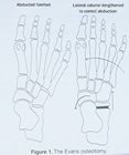 Procedure is used to correct abduction of the forefoot. Abduction is a transverse plane deformity. The test used to determine the amount of abduction present in the forefoot is called a "too many toes" sign. In cases of extreme forefoot abduction, when the foot is viewed from the back, the fourth and fifth toes will be seen peeking out along the lateral aspect of the foot. The Evans Procedure is used to wedge the foot back to a straight, or non-abducted position. An Evans Procedure uses a bone graft to wedge the distal calcaneus, in effect lengthening the lateral column of the foot. An Evans Procedure may be performed in conjunction with other flatfoot procedures.
Rigid flatfeet also are treated with tendon transfers. The most common tendon transfer in flatfeet surgery is the transfer of the flexor hallucis longus tendon to the posterior tibial tendon.(16) The posterior tibial tendon is the primary tendinous support of the medial arch. The posterior tibial tendon often fails in cases of flatfeet. Tendon transfers such as this serve to reinforce the PT tendon.
Treatment of rigid flatfeet deformity can be challenging for both the surgeon and patient. When planning rigid flatfeet correction it's important that patients understand the degree of disability associated with the procedure. It is not unusual for many patients to be off work for six months or more.
When to contact your doctor
Many problems with pediatric and adult flatfeet can be addressed with the use of an arch cookie, OTC arch support or shoe modifications. If a flatfoot remains symptomatic following a reasonable period of conservative care, consult your podiatrist or orthopedist for additional treatment recommendations.
References
1. Bordelon RL. Correction of hypermobile flatfoot in children by molded insert. Foot Ankle 1:43-150, 1980.
2. Wenger DR, Mauldin D, Speck G, Morgan D, Leiber RL. Corrective shoes and inserts as treatment for flexible flatfoot in infants and children. J Bone Joint Surg Am 71:800-810, 1989.
3. Bowman GD. New concepts in orthotic management of adult hyperpronated foot: preliminary findings. J Prosthet Ortho 9:77-81, 1997.
5. Giannini S, Kenneth A, Johnson Memorial Lecture. Operative treatment of the flatfoot: why and how. Foot Ankle Int 19:52-58, 1998.
6. Giannini, BS, Ceccarelli F, Benedetti MG, Catani F, Faldini C. Surgical treatment of flexible flatfoot in children a four year follow-up study. J Bone Joint Surg Am 82-A:73-79, 2008.
7. Vogler, HW Arthroeresis principles and concepts. In: Yearbook of Podiatric Medicine and Surgery, p. 448, edited by T Clark, Futura, Mt Kisco, NY, 1981.
8. Cantanzariti AR, Mendicino RW, Saltrick KR, Orsini RC, Dombek MF, Lamm BM. Subtalar joint arthroeresis. J Am Podiatr Med Assoc 95:34-41, 2005.
9. Easley ME, Trnka HJ, Schon LC, Myerson MS. Isolated subtalar arthroeresis. J Bone Joint Surg Am 82:613-624, 2000.
10. Kitaoka HB, Patzer GL, Subtalar arthroeresis for posterior tibial tendon dysfunction and pes planus. Clin Orthop Relat Res Dec: 187-194, 1997.
11. Lanham RH. Indications and complications of arthroeresis in hypermobile flatoot. J Am Podiatry Assoc 69:178-185, 1979.
12. Scharer BM, Black BE, Sockrider N. Treatmetn of painful pediatric flatfoot with Maxwell-Brancheau subtalar arthroeresis implant a retrospective radiographic review. Foot Ankle Spec 3:67-72, 2010.
13. Koning PM, Heesterbeek PJC, de Visser E. Subtalar arthroeresis for pediatric flexible pes plano valgus: fifteen years experience with the cone-shaped implant. J Am Podiatri Med Assoc 99:447-453, 2009.
14. Lepow GM, Smith SD, A modified subtalar arthroeresis implant for the correction of flexible flatfoot in children. The STA Peg procedure. Clin Podiatri Med Surg 6:585-590, 1989.
15. Vora AM, Tien TR, Parks BG, Schon LC. correction of moderate and severe flexible flatfoot with medializing calcaneal osteotomy and flexor digitorum transfer. J BoneJoint Surg Am 88:1726-1734, 2006.
16. Cicchinelli LD, Huerta JP, Carmona FJG, Morato DF. Analysis of gastroc recession and medial column procedure as adjuncts in arthroeresis for the correction of pediatric pes planovalgus: a radiographic retrospective study. J Foot Ankle Surg 47:385-391, 2008.
Author(s) and date
![]() This article was written by Myfootshop.com medical advisor Jeffrey A. Oster, DPM.
This article was written by Myfootshop.com medical advisor Jeffrey A. Oster, DPM.
Competing Interests - None
Cite this article as: Oster, Jeffrey. Flatfeet https://myfootshop.com/article/flatfeet
Most recent article update: December 10, 2020.
 Flatfeet by Myfootshop.com is licensed under a Creative Commons Attribution-NonCommercial 3.0 Unported License.
